Deaf mother, athlete, and teacher of the deaf
December 21, 2022Top Hearing Like Me Posts of 2022
December 30, 2022Researchers prove accessibility of the cochlea
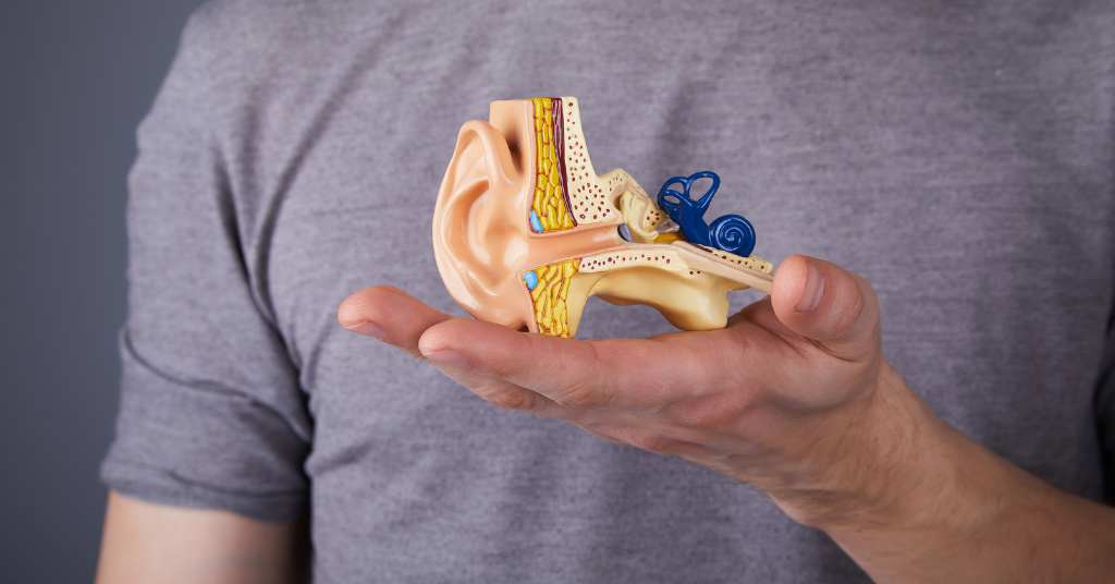
Often referred to as a snail shaped structure, the bony cochlea safely encloses our most precious hearing capabilities. Tucked inside are thousands of hair cells that transmit electrical wavelengths to the brain. Once they reach the brain, they are comprehended as the sounds one enjoys. A new study is debunking previously believed notions about accessibility to this part of the ear.
Discovering a Safe Passageway Into the Inner Ear
Though intensely studied, according to Radifah Kabir in an ABP Live article, the cochlea seemed to be inaccessible in years past. However, researchers from across the world came together to disprove this. The team consisted of Guy’s and St. Thomas’ NHS Foundation Trust in London, Uppsala University in Sweden, University of Sheffield in UK, University of Nottingham in UK, and Western University in Canada. Using advanced imaging technology, including temporal bone models, the researchers had exciting results.
Their published paper can be found in the Scientific Reports Journal, titled, “Unlocking the human inner ear for therapeutic intervention.” The sponsor of this study is Rinri Therapeutics, a group whose mission is discovering regenerative cell therapy to restore hearing. They found that the pathway into the cochlea is known as the Rosenthal’s canal. Rosenthal’s canal has a diameter of .1 to .05 mm. Inside this canal are auditory neurons. As one can imagine, this is an extremely tiny passageway. Although small in size, the team discovered that this canal may hold the secret to realistic access to the inner ear. With this newfound, safe access, researchers throughout the globe are encouraged to continue looking into gene therapies and regeneration possibilities.
With this newfound, safe access, researchers throughout the globe are encouraged to continue looking into gene therapies and regeneration possibilities.
Read more: How the Cochlea Contributes to Hearing
Methods
How did the researchers analyze such a narrow opening? How was safety considered? Researchers started by using specialized imaging technology called synchrotron radiation phase-contrast imaging (SR-PCl). This SR-PCl allows for the high-definition images to distinguish the edges of the soft tissue more accurately from the bone.
To assess safety, computer vision technology enabled the researchers to visualize the blood vessels and nerve pathways. This specialized technology enabled the team to gauge the risk of potential harm. These imaging studies allowed them to create an anatomically correct human inner ear. The fine-tuned accuracy of these imaging tools has enabled researchers and physicians to better understand the structure of the inner ear. Research and clinical practices continue to advance.
Read more: Unlocking the human inner ear for therapeutic intervention
Implications of This Research
Sensorineural hearing loss is steadily on the rise. According to the World Health Organization, it is estimated that about 700 million people will have hearing loss by 2050. Additionally, hearing loss can be caused by a variety factors including genetics, congenital conditions, infections, listening to loud sounds, etc. Due to the high prediction of increased hearing damage, there are goals to better both hearing technology and cell regeneration therapies.
This latest research is valuable as it shows an applicable access point of the cochlea. In a News Medical Life Sciences article, Professor Marcelo Rivolta explains why this is such a big deal: “Until now this region of the inner ear has been inaccessible in humans. This means that the pioneering advanced therapies to repair the auditory nerve, which have already proved successful in animal models, have been hampered by limited anatomical knowledge and the lack of a safe access to Rosenthal’s canal – the compartment that houses the auditory neurons within the central core of the cochlea.”